
 Request an Appointment
.webp?sfvrsn=276ce14_1/vector-(3).webp) International Patient
International Patient

.webp)

Breast cancer is a cancer arising from the uncontrollable cell growth of cells in the breast. It refers to cancer originating from breast tissue, most commonly from the inner lining of milk-producing ducts (such cancers are known as “ductal carcinoma”) or the lobules that supply the ducts with milk (also known as “lobular carcinoma”).
What are the types of breast cancer?
Breast cancer is classified by its location of origin and whether it spreads from the origin.
Non-invasive breast cancer is cancer of the cells of the breast duct or lobule (milk glands), which does not spread from this location.
Ductal carcinoma in situ (DCIS)
DCIS is a precancerous condition where abnormal cells are found in the lining of the breast milk duct. This condition is non-invasive and thus, the abnormal cells have not spread to the surrounding tissue. DCIS is the earliest form of breast cancer and is commonly discovered during a mammogram. Although it is non-invasive, DCIS still requires treatment to ensure that it does not spread to surrounding breast tissue.
Lobular carcinoma in situ (LCIS)
LCIS is a condition where abnormal cells are found in the lobules (milk glands) of the breast, which have not spread to the surrounding tissue. While this condition is not yet cancer, it means you do have an increased risk of getting breast cancer.
Invasive breast cancer is cancer of the cells of the breast duct or lobule that spreads and invades surrounding tissue. It can also spread to other regions of the body through the blood and lymphatic systems.
Invasive ductal carcinoma (IDC)
Abnormal cells in this cancer started in the milk ducts and have spread to the surrounding tissue.
Invasive lobular carcinoma (ILC)
This cancer begins in the lobules, or milk-producing glands of a woman’s breast. The term invasive in its name means that the cancerous cells which started in the lobules of the breast have spread to the nearby lymph nodes and maybe to other parts of the body.
Can men get breast cancer?
Men can also get breast cancer, although it is rare. Although men’s breasts are not fully developed like women’s, men too have breast tissue. Cancerous cells can also form in male breast tissue and are more common in older men.
Below are the most common types of breast cancer in men:
- Invasive ductal carcinoma (IDC)
- Invasive lobular carcinoma (ILC)
- Ductal carcinoma in situ (DCIS)
Men with breast cancer usually have a good prognosis if the disease is detected early.
What is triple negative breast cancer?
Triple negative breast cancer refers to cancers that test negative for oestrogen, progesterone and the HER2 protein. This type of cancer is usually more aggressive and as such, very likely to have spread to surrounding areas by the time it is detected.
Triple negative breast cancer presents with the usual breast cancer symptoms, such as a lump in the breast, redness or pain in the breast, and a nipple that has discharge or turns inward.
What are the stages of breast cancer?
What are the signs and symptoms of breast cancer?
The symptoms of breast cancer vary widely. Some types of breast cancer may not show obvious symptoms. Some warning signs of breast cancer are:
Skin changes such as redness, discolouration, scaliness or mild flaking of the nipple skin could be due to Paget’s disease of the breast, one of the many variants of breast cancer.
Irritation or dimpling of the breast skin may occur as well while in inflammatory breast cancer; the skin may have ridges or appear pitted, akin to the skin of an orange (called peau d’orange).
Signs and symptoms also include thickening or swelling of a part of the breast and change in breast size, shape, or appearance.
Some changes with the nipple are trivial, such as structural (retracted or inverted), while others may occur due to the presence of discharge (clear or bloody discharge) that is not breast milk.
Note that the above symptoms can arise with other conditions that are not cancer. If you feel concerned over certain signs or symptoms, see a doctor right away.
In advance stages, symptoms related to distant spread of the cancer may occur, such as:
- Bone pain (bone metastases)
- Shortness of breath (lung metastases)
- Drop in appetite (liver metastases)
- Involuntary weight loss (liver metastases)
- Headaches
- Neurological pain or weakness
What are the risk factors of developing breast cancer?
There are certain risk factors that you should be aware of when it comes to breast cancer causes. These include:
- Female gender (breast cancer is far more common in females)
- Family history of breast cancer
- Carrier of BRCA gene mutations (BRCA 1 and BRCA 2)
- Previous history of breast cancer
- Presence of breast lumps
- Having a first child at an older age
- Never having been pregnant (never conceived)
- Use of hormonal replacement therapy
- Alcohol and cigarette use
- Older age
- Radiation exposure (from radiation therapy treatments)
- Started menstruation at a younger age (before 12 years old)
- Menopause at an older age (after 55 years old)
- Being overweight or obese
How do doctors diagnose breast cancer?
Breast cancer is generally diagnosed either through pre-empted screening or diagnostic test prompted by its symptoms (e.g., breast pain, nipple discharge or presence of mass). Regular health screenings are important as they allow early detection of tumours. When cancers are detected early, they tend to have better treatment outcomes.
Patients who present breast cancer symptoms will undergo a set of assessments known as ‘Assessment’. The Triple Assessment test is used to accurately diagnose all palpable breast lumps and can yield a sensitivity of 99%.
Triple Assessment consists of:
Doctor’s assessments include findings from clinical breast examination (CBE). A doctor will conduct CBE to notice any change involving the size, shape, and texture of any lump.
Benign lumps are soft, smooth, round, and movable. A malignant lump feels hard, is oddly shaped, and feels firmly attached.
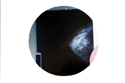
A mammogram is an x-ray of the breast. Mammograms can detect breast cancer years before the tumour can be felt by you or your doctor. A mammogram should be scheduled every year after age 40.
How does mammography screening help detect breast cancer?
- Mammograms can detect breast lesions smaller than 1 cm in size.
- A smaller lesion increases the possibility of having breast-conserving surgery.
- Survival rate is also generally better.
A breast ultrasound looks inside of the breast using sound waves and their echoes. It can detect certain features that may be difficult to observe on mammograms, such as fluid-filled cysts.
How does an ultrasound help detect breast cancer?
- Ultrasounds are useful to distinguish solid tumours from fluid-filled masses such as cysts, which are very unlikely to be cancer.
- Ultrasounds are also used to guide a biopsy needle into the breast tissue to extract cells and examine them for cancer.
A breast biopsy may be performed if symptoms or an imaging test (such a mammogram) indicate the possibility of breast cancer. A small amount of breast tissue is taken from the suspected lesion so they may be tested in a lab.
How does a biopsy screen for breast cancer?
- A biopsy allows for tissue samples from the breast to be examined for the presence of cancer cells.
- It is performed to confirm a cancer diagnosis.
Biopsy of breast tissues can be obtained either through fine needle aspiration cytology (FNAC) or core needle biopsy (CNB):
- FNAC is the least invasive method of breast biopsy which involves the insertion of a thin, hollow needle into the breast to withdraw cells from the suspected area.
- CNB uses a larger needle and instead of cells, it removes a small cylinder of tissue (a core) about the size of a rice grain, from the breast.
From the breast biopsy, pathologists can determine the appearance of cells, grading and cancer stage.
Learn more about the different types of screening and diagnostic procedures performed to diagnose breast cancer.
How is breast cancer treated?
Management of breast cancer depends on several factors such as the stage of the cancer, tumour-to-breast ratio, and the patient’s condition.
After the diagnosis has been established, the doctor will provide a comprehensive counselling on the treatment options based on their respective outcomes. Patients would then be able to make an informed decision regarding the best treatment for themselves.
Treatment for breast cancer depends on the size of the tumour and the extent of its spread in the body. These may include:
Mastectomy involves the complete removal of the breast, and it is performed under the following considerations:
- Factors that increase the risk of local recurrence (e.g., diffuse visible calcifications on mammogram, multiple primary tumours, or failure to obtain tumour-free margins).
- Physical disabilities that preclude lying flat or abducting the arm, preventing the use of radiotherapy.
- Absolute contraindications to radiotherapy such as pregnancy, previous irradiation of the breast or relative contraindications such as systemic lupus erythematosus or scleroderma.
- Tumour of 4 cm or more in size, or larger tumours with sizes in proportion to the breast. Neoadjuvant (i.e., pre-surgery) chemotherapy has the potential to downstage large tumours and improve the chances of successful breast conserving surgery.
- The patient’s clear preference for mastectomy.
Chemotherapy is a systemic therapy that can either be performed as an adjuvant or neoadjuvant therapy (before a surgical procedure). Neoadjuvant chemotherapy is usually provided for locally advanced breast cancer.
As breast cancer is recognised as a systemic disease even in its early stage, adjuvant chemotherapy should be considered, especially in pre-menopausal women who have the following:
- One or more positive axillary lymph nodes
- Oestrogen receptor (ER) negative disease
- HER2, 3+ disease
- Tumour larger than 2 cm in size
- Grade 3 disease
Neoadjuvant chemotherapy is also increasingly being used for certain patients with earlier stage disease (in particular, patients with triple negative disease or HER2-positive disease).
Monoclonal antibodies can also be used to treat breast cancer. Normally, the body creates antibodies in response to an antigen (the receptor on the surface of bacteria). Antibody attaches to the antigen in order to mark it for destruction by the body’s immune system. Trastuzumab (a monoclonal antibody) works by targeting receptors on cancer cells.
Under regular conditions, the human epidermal growth factor receptor 2 (HER2) gene stimulates normal cell growth by instructing the cell to divide and multiply in a controlled manner. However, some cancerous breast tissues contain too much HER2, triggering the cells to divide and multiply rapidly. Trastuzumab attaches to the HER2 receptors and marks the cell for the immune system to destroy.
Endocrine therapy is beneficial for patients who suffer from oestrogen receptor (ER) positive diseases. Treatments under this group of therapy include the use of systemic drugs such as tamoxifen and aromatase inhibitors, and ovarian suppression or ablation therapy.
Learn more about the different types of treatment technologies to treat breast cancer.
What can I do to reduce the risk of developing breast cancer?
We recommend following these healthy lifestyle practices to reduce the risk of breast cancer:
- Maintaining a healthy body weight
- Exercise regularly
- Eat a well-balanced, healthy diet
- Avoid or stop smoking
- Avoid or limit alcohol
- Do breast self-examination (breast check) frequently
- Limit postmenopausal hormone therapy
Frequently asked questions (FAQs)
Risk factors
Yes, breastfeeding can reduce the risk of developing breast cancer. Hormonal changes after giving birth pause a woman’s period. Breastfeeding extends these changes, reducing exposure to hormones such as oestrogen over her lifetime. Since these hormones are linked to breast cancer, breastfeeding reduces the risk of breast cancer.
Besides this, women who breastfeed shed breast tissue as they do so. This process can help remove cells with DNA damage and thus, reduce the chances of contracting breast cancer.
Signs & symptoms
Cancerous lumps are usually:
- Painless
- Feels like a hard mass
- Immobile - does not move around when pushed
- Irregular in shape
- Located at the outer part of your breast
- Grows over time
However, not all cancerous lumps feel this way. They may also be soft, tender, rounded and painful. As such, it is imperative that you see a doctor right away if you feel any lump.
Treatment
Early detection & prevention
Women should perform a breast self-examination (BSE) regularly starting from age 20. Conduct a breast self-exam once a month and look for any changes in breast tissue, such as changes in size.
If you notice any palpable lumps, dimpling or puckering of the breast, inversion of the nipple, redness or scaliness of the breast skin, nipple/areola area, or discharge of secretions from the nipple, consult your doctor without delay.
How to do breast self-examination (breast check)

1. Stand in front of a mirror and look at your breasts closely. Look for changes in size, shape, colour and texture of your skin and nipple.
2. Next, lift your arms above your head and check your breasts again.
3. Then, place your hands on your hips and press inwards to tense the muscle under your breasts.
4. Look closely for any visible changes in the breasts.

5. Begin palpation from your underarm and examine your breast in concentric circles, starting from the outer region of the nipple.
6. Your breasts can also be palpated in vertical strips.
7. Gently squeeze your nipples to look for abnormal discharge.
8. With your arm placed at the side, examine for lumps under your armpit again using your free hand.
Changes in your lifestyle can help decrease the risk of breast cancer. You can do the following:
- Consult your doctor about breast screening
- Be familiar with your breasts and inspect for changes, lumps, or other signs. While this can’t prevent breast cancer, it will help you to catch it early on
- Limit alcohol intake
- Exercise 30 minutes a day
- Be aware of what you eat. A healthy diet can reduce your risk of breast cancer
- Maintain a healthy weight
- Be aware of the risks of postmenopausal hormone therapy
Myths & facts
Make an appointment at Pantai Hospitals
Early detection is important for the best outcomes. Performing regular breast self-examinations and scheduling regular mammograms can help detect breast cancer in its early stages. Early-stage detection gives you the best chance of fighting breast cancer.
Early detection of breast cancer makes it easier to treat the disease with effective and appropriate treatment. A dedicated multidisciplinary team of specialists and oncologists at Pantai Hospitals is available for consultation to provide the best care and assistance to patients through screening, diagnosis, and treatment.
Get in touch with us to book an appointment today if you have any concerns or questions about breast cancer treatment options.
Learn more about cancer care and Oncology services at Pantai Hospitals.
Pantai Hospitals have been accredited by the Malaysian Society for Quality in Health (MSQH) for its commitment to patient safety and service quality.

.jpg?sfvrsn=2a4fde48_1)

.png?sfvrsn=b7ea172b_1)
 Find A Doctor


.webp?sfvrsn=20763f7d_21)
.webp?sfvrsn=f2a2c343_12)