.webp)
Screening is getting checked for cancer when you do not show any symptoms. Regular cancer screening increases the chances of early cancer detection; early-stage cancer is also easier to treat. Taking a proactive stance and getting yourself screened can help protect your well-being.
There are various screening tests to detect a few common cancers found in Malaysia. Your doctor would recommend the best screening tests for you, based on your risk factors.
Mammography
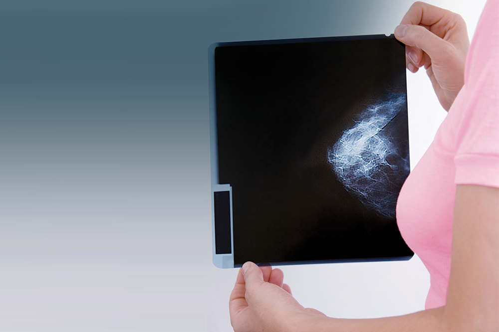
A mammogram is an X-ray of the breasts. The X-ray image taken is examined for signs of cancer. This procedure is a useful breast cancer screening tool to detect the presence of small lumps in the breast that cannot be felt by hand.
Digital mammography uses a low dose of radiation and lesser breast compression. It gives accurate results and more detailed depiction as compared to conventional (analogue) mammogram.
Who should go for mammography?
Book a mammography appointment at the Pantai Hospital nearest to you.
Pap test
The Pap smear test (or Pap test) is a screening test to detect abnormal cell changes of the cervix (the lower part of the uterus that opens into the vagina). The doctor will examine the vagina and cervix and takes some samples to be sent to a lab for testing. This helps detect abnormal cells before they become cancerous.
HPV test
The HPV test looks for the presence of the human papilloma virus, which increases your risk of cervical cancer. An HPV test can be performed at the same time as a Pap test. The doctor will insert a speculum to push apart your vaginal wall, followed by a spatula to collect cells from your cervix. These cells will then be sent to the laboratory for testing.
Who should go for Pap test and HPV test?
Book a pap test and HPV test appointment with a gynaecological oncology specialist at the Pantai Hospital nearest to you.
Faecal Occult Blood Test (FOBT)
Faecal occult blood test (FOBT) is a lab test that can detect blood in stool that are not seen by the naked eye. The stool is collected at home and sent to a lab. Individuals above the age of 50 are encouraged to do FOBT once a year.
Flexible sigmoidoscopy
A short tube is inserted into the rectum to observe for polyps or cancerous masses in the rectum and lower colon.
Colonoscopy
A longer tube is inserted into the rectum to observe for polyps or cancerous masses in the rectum and entire colon. A camera embedded at the end of a flexible tube is passed through the anus, allowing for examination of the large bowel and part of the small bowel. Polyps can be removed during colonoscopy as well.
CT colonography
X-rays and computers are used to produce images of your entire colon to observe for abnormal growths.
Each test differs in its advantages and disadvantages. Your doctor would recommend the most suitable test for you.
Who should go for colorectal (colon) cancer screening?
Book an appointment with a gastroenterologist at the nearest Pantai Hospital to you.
Low-dose computed tomography (LDCT)
During the scan, you will be asked to lie down on a table. A computer linked to an X-ray machine will produce a series of detailed images of your lungs to be examined by your doctor.
Who should go for yearly lung cancer screening with LDCT?
*One pack-year is smoking an average of one pack of cigarettes per day for one year. A 20 pack-year smoking history translates to smoking one pack a day for 20 years, or two packs a day for 10 years, and so on.
Book an appointment with a respiratory physician at the Pantai Hospital nearest to you.
If you would like to find out more about screening procedures for other types of cancers, please refer to the individual cancer articles available by accessing the interactive human body map for cancer on our Oncology (Cancer) website.
Find out more about cancer screening packages available at the Pantai Hospital nearest to you.
There is also a wide range of health screening programmes offered by Pantai Hospitals. They are specifically designed and can be customised based on your medical needs. To book an appointment, please contact the health screening centre at the nearest Pantai Hospital to you.
A cancer diagnosis is made after performing various tests. Your doctor would ask about the history of your health and symptoms. Then, your doctor would perform at least one of the examinations below.
Abnormal levels of certain substances in the body can indicate cancer. Laboratory tests help to detect changes in the amount of these substances. These tests include urine test, blood test, and tumour marker test.
Tumour markers, for example, are produced by cancer cells or by other cells in response to cancer. Therefore, they can be used to diagnose cancer. Common tumour markers are:
A biopsy may be performed in most patients to confirm the cancer. In a biopsy, the doctor takes a sample of cells from the suspected mass. These cells are viewed under a microscope and may undergo some tests.
A biopsy may be carried out in several ways:
Imaging tests are non-invasive procedures that produce images of inside the body, so your doctor may check for presence of tumours. Examples of imaging tests include X-ray, ultrasound, Computerised Tomography (CT) scan, Positron Emission Tomography (PET) scan, and Magnetic Resonance Imaging (MRI).
Digital Radiography (X-ray)

Digital radiography (X-ray) utilises a low dose of radiation to capture images of inside the body. Read more
A technician will assist you to position your body so that the X-ray can be directed at the target part of your body and the image is captured for your doctor to view them. Due to lower doses of radiation, digital x-ray minimises damage to cells in your body.
Ultrasound
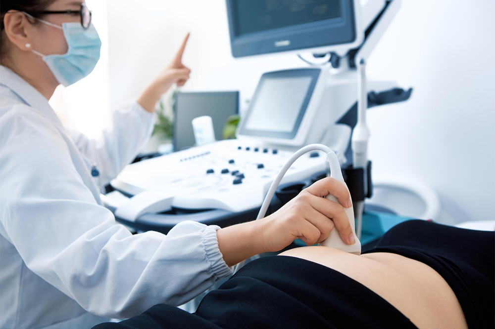
Ultrasound uses sound waves or frequency that reflect off the tissues in the body to create images on a computer. Read more
The sound waves will bounce off tissues in your body and produce echoes that are used by a computer to create images in real-time. These echo waves will be measured to determine the object’s distance, size, shape, and consistency (solid, filled with fluid or both).
During an ultrasound procedure, you will be asked to lie down. A device called a transducer is lubricated with gel and glided on the skin over the area being examined.
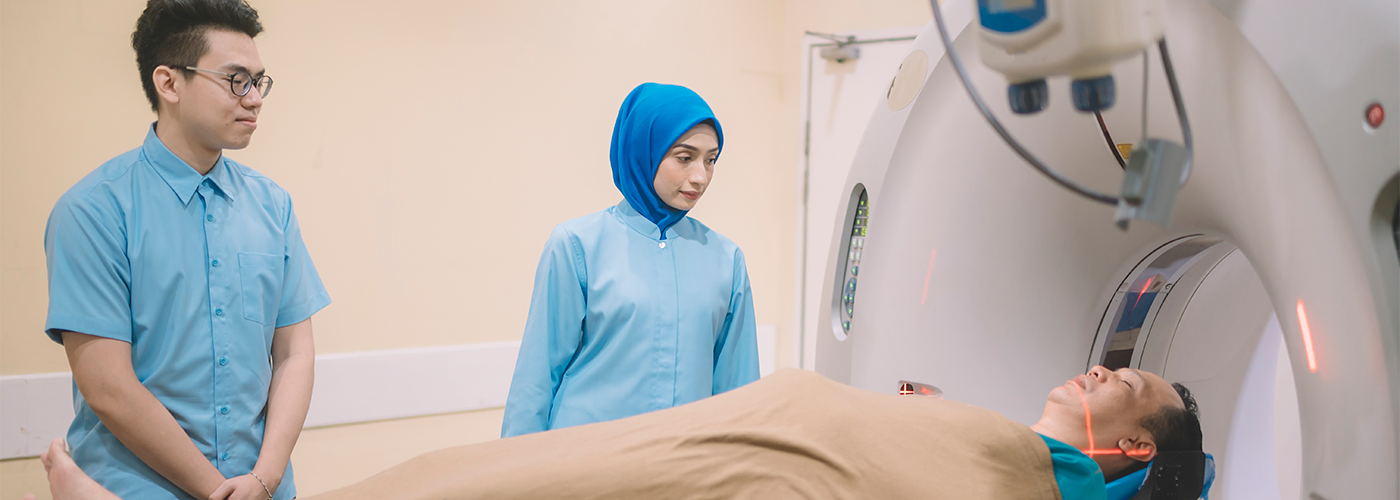
Computed Tomography (CT) Scan
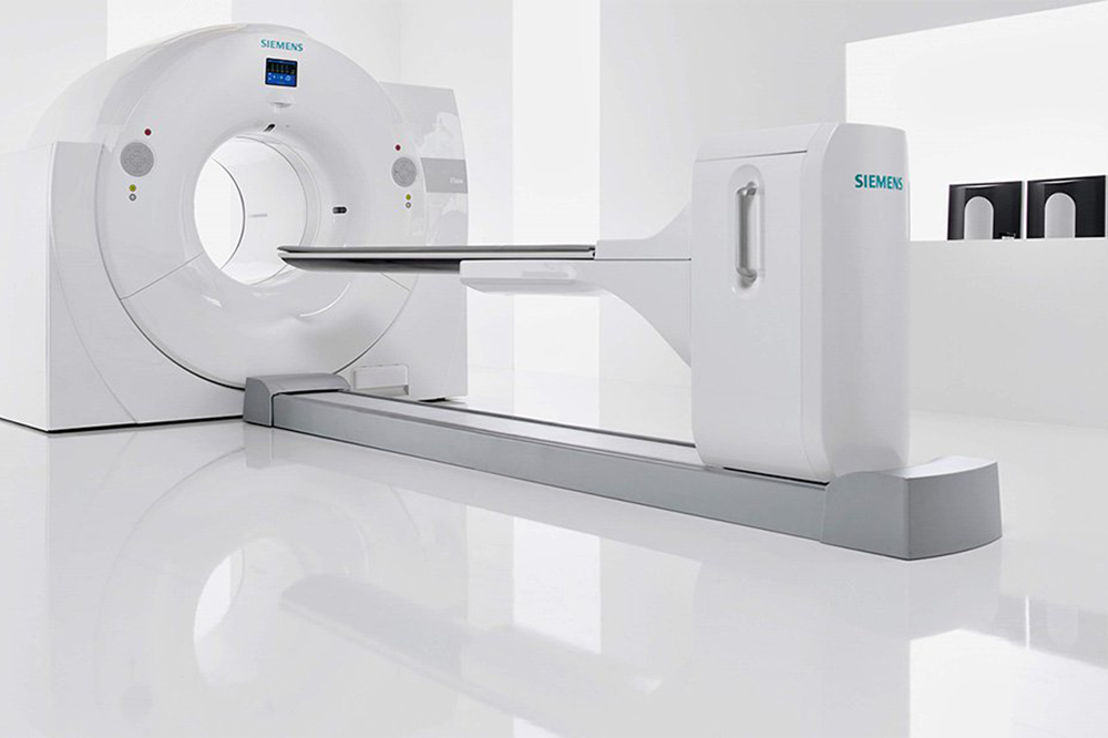
A Computed Tomography (CT) scan is a non-invasive imaging test that combines X-rays and computers to generate detailed anatomical images (slices) of bones, blood vessels, and soft tissues in your body. Read more
Recent advances in Computed Tomography (CT) technology have improved the quality of imaging by providing fast, thin-slice, and wide-range scanning capability. Up to 31% less radiation dose could be used during imaging, thus minimising radiation exposure for patients.
During a CT scan procedure, you may be given an injection or oral contrast that helps make the images clearer if your attending doctor requests for a CT scan with contrast.
Then, you would be asked to lie down on a table that slides you into a doughnut-shaped machine for the images to be taken. You will not feel anything during the scan. The scan may take 15-30 minutes depending on the body part being examined.
The 640-slice CT Scanner is equipped with VISION Optics that allows the x-ray spectrum to be optimised for low dose and high-quality images scanning. It can generate precise images of soft tissues, bones, and blood vessels, thus leading to more accurate diagnosis.
Magnetic Resonance Imaging (MRI) Scan
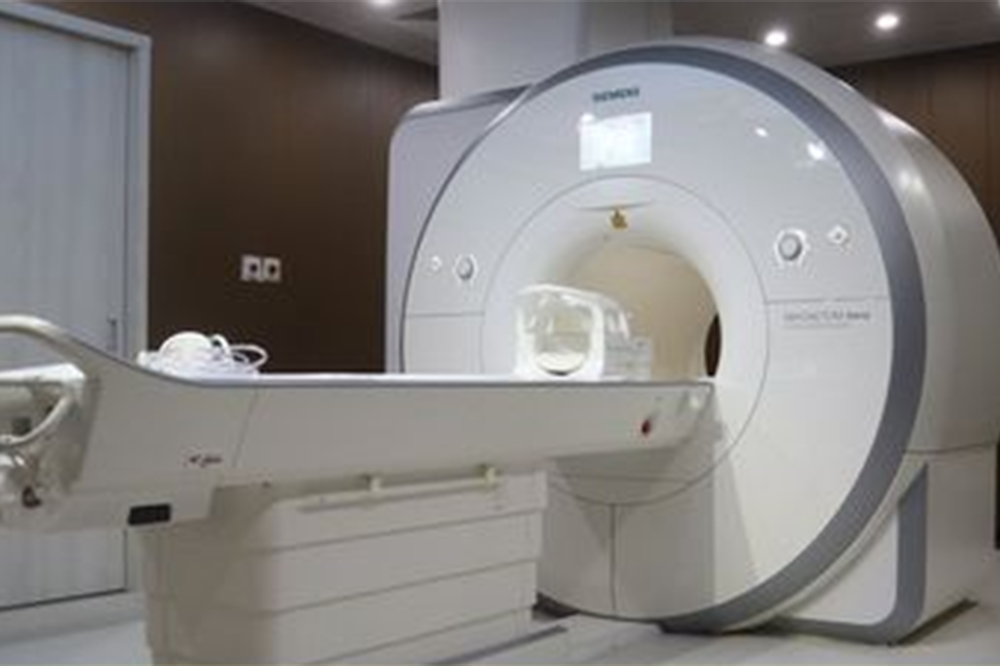
Magnetic resonance imaging (MRI) scan uses powerful magnetic and radio waves to produce detailed images of the inside of your body. It is a non-invasive imaging test without exposure to radiation (X-rays). Read more
MRI scans are very useful for viewing soft tissues of the body such as the heart, lungs, liver, or other organs. MRI scans produce better images of the soft tissues than X-rays. Doctors would then study the images to identify, diagnose, and evaluate various medical conditions that affect soft tissues. For example, MRI scans are able to diagnose the type of brain lesion when looking at brain tumours.
During an MRI, you would be asked to lie down in a long, round chamber. You may also receive an injection of a type of dye to create clearer images of the tumour.
The 1.5 Tesla Siemens Avanto MRI Scanner system provides comfort to the patient during the scan and produces better imaging results for the clinicians.
Single Photon Emission Computed Tomography (SPECT)
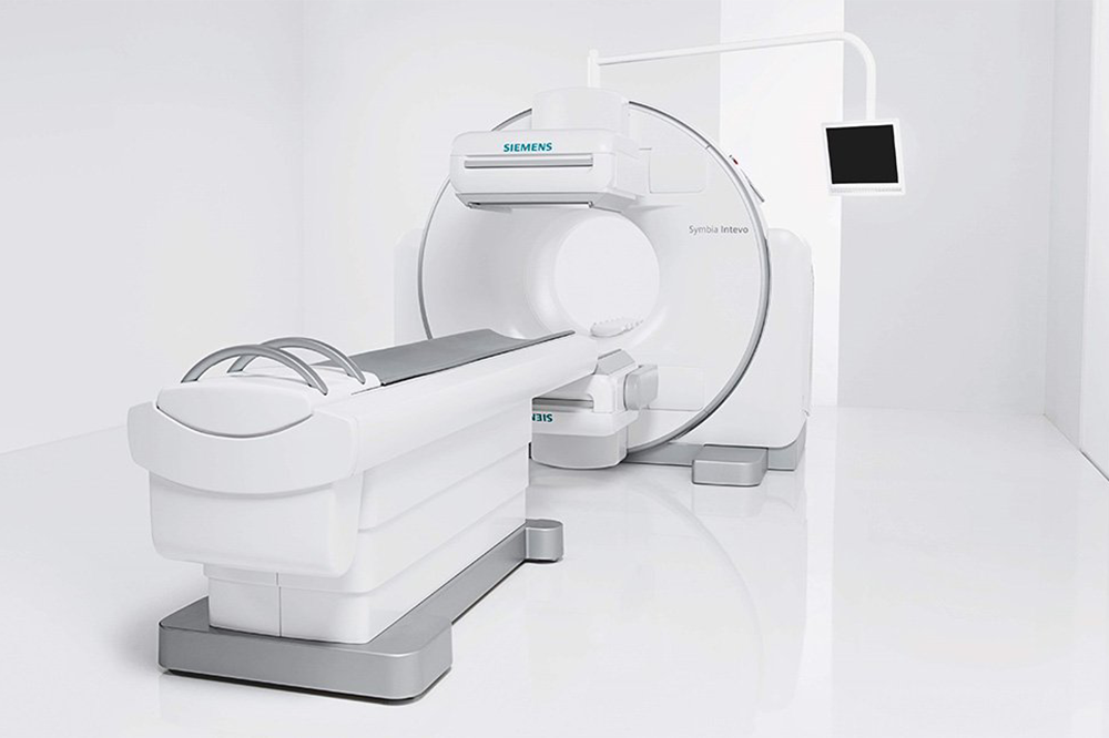
Single Photon Emission Computed Tomography (SPECT) is a non-invasive, advanced nuclear imaging test used frequently in diagnostic medicine. Read more
SPECT-CT scan
SPECT scans can show biological functions of organs and systems through the distribution of radiotracers in the body. Combined with CT technology, the SPECT-CT modality can identify precise location and anatomical structures.
SPECT-CT technology has become a superior diagnostic imaging modality used in diagnosis and management of cancer and other diseases.
The Siemens Symbia Intevo T16 SPECT/CT system combines Single Photon Emission Computed Tomography (SPECT) with high quality 16 slices diagnostic Computed Tomography (CT) to generate higher SPECT images resolution and accurate, reproducible quantitative results. This machine can operate as SPECT-only, SPECT-CT or stand-alone CT diagnostic application.
Learn more about the Siemens Symbia Intevo T16 SPECT/CT here.
Positron Emission Tomography (PET) Scan
Positron Emission Tomography (PET) is a non-invasive, advanced nuclear diagnostic scan that provides detailed information on the activity of an organ or a system in your body.Read more
PET-CT scan
A Positron-Emission Tomography (PET) scan can be combined with a Computed Tomography (CT) scan. This is called a PET-CT scan that combines the functional imaging (showing the activity of an organ or system in your body) capability of a PET with the anatomical imaging (detailed pictures of organs) of a CT.
PET-CT is commonly used in cancer diagnosis due to its capability of improving the accuracy of cancer staging, helping with planning for biopsy and radiotherapy, and allowing the observation of new cancer growth after treatment. Therefore, PET-CTs have become the best modality in monitoring the progress of cancer treatment.
During the PET-CT scan, you would be injected with a tracer (radioactive glucose). A camera detects the radioactive glucose in your body to form images. Cancer cells take up more glucose than normal cells, so this is how the PET-CT scan is used to detect cancer.
The Siemens Biograph mCT S 64 PET-CT system utilises fluorodeoxyglucose (F-18 FDG) to visualise the molecular function of active cancer cells and their spread throughout the whole body. This machine also utilises radiopharmaceutical tracer to perform PSMA PET-CT Scan for prostate cancer and Gallium-68 DOTATATE PET-CT Scan for neuroendocrine cancer.
Learn more about the Siemens Biograph mCT S 64 PET-CT here.
If you experience any signs and symptoms of cancer or would like to be screened for cancer, get in touch with us to find out more about our Oncology Services at your nearest Pantai Hospital.
A dedicated multidisciplinary team of specialists and oncologists at Pantai Hospitals is available for consultation to provide the best care and assistance to patients through screening, diagnosis, and treatment.
Pantai Hospitals have been accredited by the Malaysian Society for Quality in Health (MSQH) for its commitment to patient safety and service quality.


.webp?sfvrsn=20763f7d_21)
.webp?sfvrsn=f2a2c343_12)

 Request an Appointment
.webp?sfvrsn=276ce14_1/vector-(3).webp) International Patient
International Patient

 Find A Doctor